Causes of Ankle Popping and Clicking
No matter your age, you might have heard a crackle or pop in your joints when sitting down, standing up or simply walking. Luckily most popping cases are not detrimental to your physical health. However, if left untreated, they could cause mobility complications.
Medically, popping in your joints is known as crepitus. If you’re experiencing a bout of crepitus, it could be from overworking your joints through exercise or stiffness in the joints after a long period of inactivity. While these causes may sound contradictory, it’s how our bodies adapt to certain stimuli — or lack thereof.
At the Orthopedic Institute of Pennsylvania, we can treat foot and ankle pain. Learn more about our services below.
Why Does My Ankle Keep Popping?
Ankle popping on its own is very common. If your popping ankle isn’t painful, it is likely caused by a gas release or tendon rubbing. However, if it is accompanied by pain or swelling, there may be an underlying cause.
The most common causes of ankle clicking or popping include:
- Gas release: Every time your ankle moves, the joint capsule filled with lubrication fluid is stretched. The fluid in the capsule can contain bubbles of nitrogen that can pop when you move, causing a loud popping sound — similar to when you pop your knuckles in your hand. Tight muscles can also contribute to gas buildup in the fluid, especially after inactivity like sleeping or binging a TV show. This is normal and does not signify an underlying cause.
- Tendon slip: The peroneal tendons in the lower outside of your leg work to stabilize the ankle joint. Sometimes, these tendons slip from the muscle surrounding them, resulting in a snapping sound or feeling. A recent ankle injury could increase the ankle popping frequency. This is also common and not a cause of concern unless it is painful.
Less common reasons that may signify an underlying health condition include:
- Tendon dislocation: The peroneal muscles surrounding the tendons can be pushed from their usual location, causing popping and snapping sounds in your ankle every time you move. This can happen during an ankle sprain. The inflammation, swelling and pain that occurs will need medical attention.
- Osteochondritis dissecans: This condition causes a small segment of bone to separate and causes the cartilage in the ankle joint to wear away. This cracking sound could be painful after a lot of movement, like walking or running.
- Osteochondral lesion: Lesions can form on the cartilage on the ends of your joint bones. Clicking and locking the ankle can occur, limiting the range of motion and causing swelling.
- Ankle osteoarthritis: Osteoarthritis refers to a joint deterioration similar to osteochondritis with the wearing down of joint cartilage over time. While most common in the knees, it can also occur in the ankle.
- Peroneal tendon injury: The peroneal tendons that may slip to make a cracking noise can also become injured.
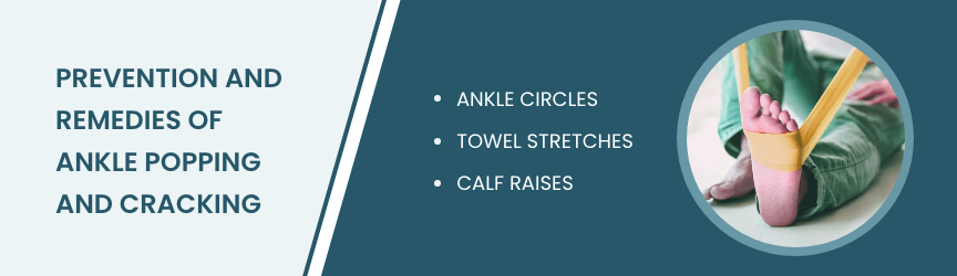
Prevention and Remedies of Ankle Popping and Cracking
You can do several exercises at home to strengthen your popping ankles. These stretches both prevent ankle popping and reduce cracking sounds that already exist.
Ankle Circles
Performing ankle circles can warm up your joints and increase mobility. You can do this exercise from a seated or lying position:
- Sit or lay with your legs stretched out front. Prop your leg on a stable surface with the ankle hanging off the edge.
- Rotate your foot in clockwise circles 10 times with your ankle.
- Switch to counterclockwise circles 10 more times.
- Swap feet and repeat with the other ankle.
Towel Stretches
Relieve tight ankles with these simple towel stretches you can do at home:
- Sit on a flat surface with your legs straight out in front of the body.
- Loop a towel horizontally around the sole.
- Gently pull the towel ends toward the body, stretching the foot.
- Hold this position for 20 seconds.
- Repeat as needed.
Calf Raises
Strengthen your calf muscles to reduce pressure on the ankles in motion.
- Stand on the edge of a platform or the bottom stair step with the heels hanging off.
- Slowly rise onto the toes, driving the body fully upward with the calves.
- Let the heels gently fall, stretching slightly below the ledge.
- Repeat 10 times.
When to See a Doctor
When your ankle popping begins to cause discomfort or pain, consider getting a proper diagnosis from a doctor. They may order tests like an MRI or CT scan to look inwardly at the bone and cartilage.
If you recently had an ankle injury, rest is a major proponent of healing. A doctor may recommend anti-inflammatory medications to help with the pain and swelling.
A doctor can stabilize the ankle with a brace or orthotic to promote faster healing for more serious conditions. Physical therapy also works alongside any stabilization devices to slowly get your ankle back to regular functioning. If necessary, surgical options are available, including arthroscopy and total joint replacements.
Visit the Orthopedic Institute of Pennsylvania Today
At the Orthopedic Institute of Pennsylvania, our ankle and foot care center is committed to helping you regain full mobility. For more information on how our services can benefit you, contact us today!


