Laminectomy

When you’re living with spinal stenosis or another spinal condition, simple joys like taking a walk, standing in line at the grocery store or sleeping through the night can become difficult. The licensed providers at Orthopedic Institute of PA (OIP) understand these challenges and will do all we can to help you regain your independence through laminectomy surgery or other proven methods.

4.9 stars 4.95 out of 5 (1611 reviews)  About star rating

Our Laminectomy Surgery Process

If your pain persists after implementing conservative treatments, we may recommend surgery as the next step. A laminectomy surgery, or decompression surgery, is intended to alleviate structural constriction affecting the spinal canal. We’ll help you understand the procedure, including how removing the lamina, the small bony arch on the back of the vertebra, can relieve some of the compression causing your symptoms. Our expert team provides support and individualized care, facilitating a comfortable path to relief.

Spinal Levels We Treat

Our team can perform a laminectomy at any level of the spine, from the occiput all the way down to the sacrum. However, we tailor each procedure to the specific level of the spine causing distress. Spinal levels are labeled L for lumbar and C for cervical. The associated number indicates the disc location. Discover how surgically treating each specific area can help:

We can also perform minimally invasive soft tissue and bone preserving laminectomies to allow for nerve decompression while minimizing trauma and complications.

Need specialized help?

We also care for many patients with other less common conditions.

Request an Appointment

Why Seek Help From OIP?

Your spine is central to everything you do, and you deserve a care team that respects that. Our patients choose us because we combine clinical excellence with genuine care. We prioritize patient education, helping you understand the how and why of the surgery. OIP experts focus on safety, long-term mobility, building relationships with our patients and offering support from initial diagnosis through recovery.

Laminectomy FAQs

Browse our frequently asked questions to learn more, or reach out to our team of skilled medical professionals to ask additional questions.

Preparation typically involves obtaining medical clearance, stopping some medications, like blood thinners, and fasting the night before your procedure.

Usually, yes. Most patients are encouraged to stand and walk with assistance within hours of the surgery to promote circulation and healing. Some patients may need longer immobilization depending on surgical findings and overall health.

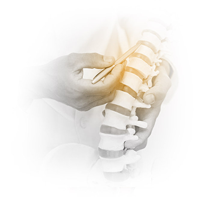
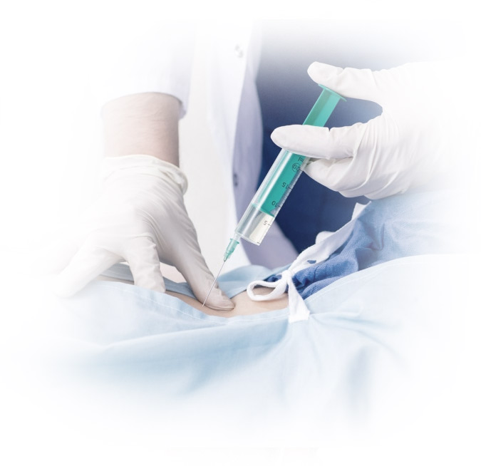
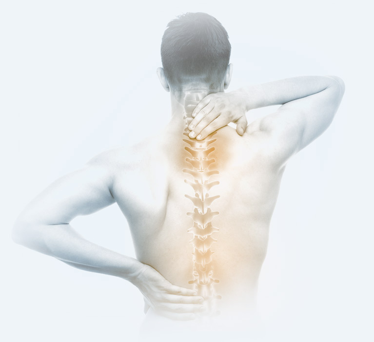
Recovery after a laminectomy usually involves a few weeks before returning to light activities and a few months before unrestricted activity. Our practice emphasizes minimally invasive spine surgery techniques that use small incisions, muscle-sparing approaches, and advanced imaging to support precision, safety, less postoperative pain, and faster recovery.

Many patients go home the same day or the next day, depending on their overall health and the extent of the surgery.

Spine Care Specialists

Patient Testimonial

After my hip replacement, I was worried about how long it would take to get back on my feet. The rehab team at OIP was fantastic. They guided me every step of the way, and I’m now walking without any pain!” Brian B., York, PA

Brian B., York, PA

MRI Upper Extremity (Shoulder, Upper Arm, Elbow, Forearm, Wrist, or Hand) Preauthorization Documentation Guidelines

1. Suspicious mass or tumor
      a. On initial evaluation or follow up
2. Staging of known cancer
3. Suspected or known infection (septic arthritis or osteomyelitis)
4. Suspected Osteonecrosis
5. Evaluation of Rheumatoid Arthritis or other autoimmune diseases
6. Evaluation of Post-op Complications
      a. Infection, delayed union, other
7. Suspected fracture with prior imaging non-diagnostic
8. Abnormal bones scan with non-diagnostic Xray
9. Significant injury with suspected ligament, cartilage, tendon, nerve or bone injury with non-diagnostic prior imaging
      a. Suspected massive rotator cuff tear
10. Evaluation of pain or more minor injury with initial imaging non-diagnostic:
      a. Pain lasting 3 months or greater
      b. Failed conservative therapy: Must include each one:
           i. Rest: modified activities or assistive devices/rigid splints or braces
           ii. Ice or heat
           iii. Medications and/or injections
           iv. Physical therapy or a physician directed home exercise program
           or chiropractic care
                1. Document instructions given
                2. Document compliance and results
                3. Document duration and dates

MRI Spine Preauthorization Documentation Criteria

1. Tumor, masses, or cancer: suspected or known
2. Neurological Deficits
3. Trauma or acute injury
       a. With neurological deficits
       b. With progressive symptoms during conservative treatment
4. Infection: known or suspected
5. Inflammation: Ankylosing Spondylitis
6. Pre-op Evaluation
7. Post-op Complications
8. Acute or Chronic Axial or radicular pain
       a. Pain lasting 6 weeks or greater
       b. ADLs must be affected
       c. Progressive neurological deficit or an abnormal EMG
             i. Must document specific dermatome, muscle weakness, reflex
abnormalities
       d. Failed conservative therapy: Must include each one:
             i. Rest: modified activities or bracing
             ii. Ice or heat
             iii. Medications, acupuncture or stimulators
                   1. Specific name of medication start date and duration and
                   results
             iv. Epidurals or other injections (not trigger point injections)
             v. Physical therapy or a physician directed home exercise program
             or chiropractic care
                   1. Document instructions given
                   2. Document compliance and results
                   3. Document duration and dates

MRI Pelvis Preauthorization Documentation Criteria

1. Musculoskeletal Pelvic MRI:
     a. Mass or tumor
    b. Significant injury to rule out fracture or other injury
    c. Osteonecrosis of hips
    d. Sacroiliitis
    e. Sacroiliac joint dysfunction
    f. Pain lasting 3 months or greater
    g. Failed conservative therapy: Must include each one:
            i. Rest: modified activities or assistive devices/rigid
            splints or braces
            ii. Ice or heat
            iii. Medications and/or injections
            iv. Physical therapy or a physician directed home
            exercise program or chiropractic care
                    1. Document instructions given
                    2. Document compliance and results
                    3. Document duration
    h. Persistent Pain not responsive to 4 weeks of conservative treatment

2. Prostate Cancer Evaluation, follow up, and surveillance

3. Mass or Tumors

4. Cancer detection, staging, or surveillance
    a. 3, 6, or 12 month follow up

5. Infection:
        a. Appendicitis
        b. Diverticulitis not responding to conservative care
        c. Inflammatory bowel disease
        d. Abscess suspected
        e. Fistula
        f. Abnormal fluid collection

6. Pelvic Floor failure

7. Uterine abnormalities

8. Undescended Testes

9. Pre-op Evaluation

10. Post-op Complication

MRI Lower Extremity (Hip, Knee, Leg, Ankle, or Foot) Preauthorization Documentation Guidelines

1. Suspicious Mass or Tumor
       a. On initial evaluation or follow up
2. Staging of known Cancer
3. Known or suspected infection
4. Suspected Osteonecrosis or Legg-Calve-Perthes Disease
5. Suspected SCFE, tarsal coaltion
6. Evaluation of Post-op complication:
       a. Infection, delayed union, other
7. Suspected fracture with prior imaging non-diagnostic
8. Abnormal bone scan with non-diagnostic xray
9. Significant injury with suspected ligament, cartilage, or bone injury
10. Evaluation of pain or more minor injury with initial imaging negative:
       a. Pain lasting 3 months or greater
       b. Failed conservative therapy: Must include each one:
             i. Rest: modified activities or assistive devices/rigid splints or braces
             ii. Ice or heat
             iii. Medications and/or injections
             iv. Physical therapy or a physician directed home exercise program orchiropractic care
                  1. Document instructions given
                   2. Document compliance and results
                   3. Document duration

MRI Chest Preauthorization Documentation Guidelines

1. Mediastinal or hilar mass
2. Myasthenia gravis with suspected thymoma
3. Brachial Plexus Dysfunction
4. Thoracic/Thoracoabdominal aneurysm
5. Suspected or confirmed Congenital Heart Disease
6. Thoracic Outlet Syndrome

MRI Brain Preauthorization Documentation Criteria

1. Suspected or known MS
2. Seizure disorder, known or suspected new or refractory
3. Suspected Parkinson’s disorder
4. Neurological symptoms or deficits
     a. Acute, new or fluctuating deficits
5. Mental status changes
6. Trauma to the head with neurological changes, vomiting, headache
7. Evaluation of headaches
      a. Chronic with change in pattern/character
      b. Sudden onset severe headache
      c. New onset headache in pregnancy
8. Suspected brain tumor or cancer
9. Known or suspected stroke
10. Suspected Infection
11. Suspected Congenital abnormality
12. New onset tinnitus or vertigo associated with visual changes

MRI Abdomen and MRCP Preauthorization Documentation Criteria

MRI
1. Suspicious Mass or Tumor
2. Surveillance of Mass, Tumor, or Cancer    
      a. 3, 6, 12 month follow up
3. Suspected infection:
      a. Appendicitis
      b. Peritonitis
      c. Pancreatitis
      d. Inflammatory bowel disease
      e. Cholecystitis
      f. Abscess
      g. Fistula
      h. Hepatitis C
4. Preoperative Evaluation
5. Post-op complication

MRCP
1. Suspected Congenital Abnormality
2. Chronic pancreatitis or related complications
3. Biliary tree symptoms
4. Pre-op Evaluation
5. Post-op complication or surveillance
6. Inconclusive abnormalities identified on other imaging